

Babies born to mothers with anaemia tend to have smaller brains than their peers, with the gap concentrated in regions tied to movement, learning and emotional regulation, according to new research. Scientists say the differences, visible from as early as age one, could set the stage for cognitive difficulties once children reach school age.
Anaemia usually caused by iron deficiency and marked by a lower-than-normal red blood cell count affects more than a third of pregnant women globally, with the highest rates in sub-Saharan Africa and south Asia. Common symptoms include fatigue, breathlessness and dizziness.
The study, led by researchers from King’s College London and the University of Cape Town, tracked more than 300 mothers and their babies in Cape Town, scanning the infants’ brains repeatedly between three months and two years of age. On average, total brain volume in babies born to anaemic mothers was 4% smaller than in babies born to mothers without the condition even though all the anaemic mothers in the study had only a mild form of it.
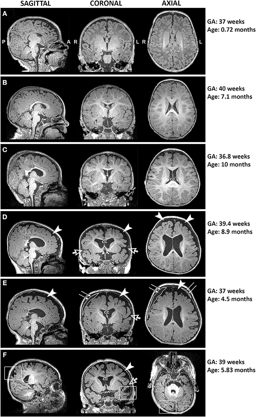
Three brain regions stood out: the putamen, the caudate nucleus and the corpus callosum. Jessica Ringshaw, the study’s first author and a researcher at King’s Institute of Psychiatry, Psychology & Neuroscience and the University of Cape Town, said these regions play a role in processing speed, emotional regulation and executive function.
The gap between the two groups widened as the children got older the corpus callosum, for example, was around 4% smaller at 12 months in babies from the anaemic-mother group, growing to a 6% difference by 24 months. That pattern echoes earlier South African research on toddlers and school-age children, which found similar brain volume differences.
Ringshaw said the gap doesn’t close over time: it persists into age six, right as children start school, which is around when subtler cognitive effects tend to become noticeable. She added that the findings help explain earlier studies showing children born to anaemic mothers performing less well on developmental tests.
According to Ringshaw, the mothers in the anaemic and non-anaemic groups were closely matched on factors like household income and food security, which gives her confidence that the anaemia itself — rather than other differences between the groups drove the brain changes observed.
The results, she said, underline why tackling anaemia in women of reproductive age matters, and she recommended routine screening for anaemia and iron deficiency during pregnancy, paired with oral or intravenous iron supplementation where needed. Anaemia’s causes are varied and can include infectious disease; the study found higher rates among women living with HIV, pointing to the need for iron interventions to go hand in hand with proper infection management.
Although the World Health Organization has named anaemia reduction a priority, global progress has largely stalled over the last 20 years. Ringshaw argued the issue needs to move higher up the public health agenda if children are to not just survive but thrive.
The research, published in the journal Brain Communications, relied on a newer generation of MRI scanners that are cheaper and more portable than standard hospital equipment. The team compared scans from conventional “high-field” machines against the portable “low-field” alternative and found the cheaper option was sensitive enough to pick up the same brain differences.
Steve Williams, professor of neuroimaging at King’s, said the study demonstrates that the technology can measure the effects of malnutrition on developing brains at scale, even in difficult settings, offering an early and sensitive way to assess the impact of planned interventions.