

A new medical ultrasound technology is opening fresh possibilities for coral conservation by allowing scientists to examine reproductive development in living corals without cutting or damaging them.
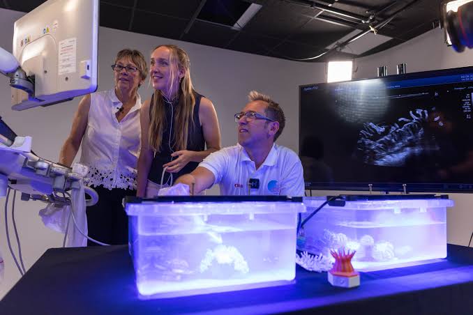
In a world-first experiment, Coral Spawning International (CSI), in partnership with Canon Medical Systems UK and Canon EMEA, used Canon’s Aplio i700 ultrasound system to identify developing eggs inside living coral, giving researchers a non-invasive way to monitor reproduction.
The breakthrough demonstrates how advanced medical imaging technology can be adapted for marine research, potentially transforming how scientists study coral reproductive cycles, predict spawning periods and support reef restoration programmes.
The experiment involved six large polyp stony (LPS) coral species with different skeletal structures, all scanned while fully submerged in water. Researchers used the Aplio i700’s high- and ultra-high-frequency probes to capture detailed images of internal coral structures and identify developing eggs.
Previously, researchers relied on invasive methods of examining coral reproduction, which involved cutting into specimens to observe their internal structures and determine whether eggs were developing.
The new ultrasound technique eliminates the need for such procedures, allowing scientists to examine living corals while preserving them for continued monitoring and research.
To verify the findings, researchers fragmented a specimen of Trachyphyllia geoffroyi and compared macro-photographs of its internal structures with the ultrasound images. The comparison confirmed that the structures identified through scanning were eggs, establishing ultrasound as a viable method of assessing coral reproductive development.
Dr Jamie Craggs, Co-Founder of Coral Spawning International, said the technology provides an unprecedented opportunity to understand the reproductive processes of coral species.
“Corals still hold so many secrets, particularly when it comes to the complex processes that drive their reproduction. Being able to look inside a living coral and see these processes unfolding in such remarkable detail is an extraordinary moment,” he said.
He added that continued monitoring would help researchers develop a more detailed understanding of how different coral species reproduce.
Sally Davey, Clinical Product Specialist at Canon Medical Systems UK, said the experiment demonstrated the versatility of ultrasound technology beyond conventional medical applications.
She noted that the successful trial could also open opportunities for applications in areas such as minimally invasive surgery and the diagnosis of dermal conditions.
Peter Bragg, Sustainability and Government Affairs Director at Canon EMEA, said the experiment highlighted the potential of imaging innovation to reveal previously unseen processes and deepen scientific understanding of the natural world.
The breakthrough comes at a critical time for coral conservation, as scientists seek better ways to understand coral reproduction and improve restoration strategies amid growing environmental pressures on marine ecosystems.
Accurately predicting coral spawning windows is particularly important because reproduction determines when corals release eggs and sperm into the water, creating opportunities for fertilisation, larval development and reef regeneration.
Following the successful experiment, the six coral species will undergo six months of monitoring, with researchers recording their reproductive development and the release of gametes.
The findings are expected to provide further insights into the reproductive cycles of LPS corals, helping scientists identify spawning periods more accurately and strengthen future coral propagation, conservation and restoration efforts.